


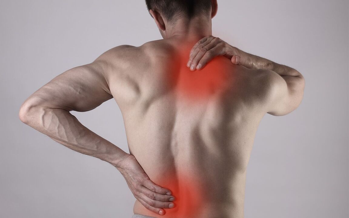
Almost every adult experiences back pain in their lifetime. This is a very common problem and can be based on various reasons, which we will analyze in this article.
Causes of Back Pain
All causes of back pain can be divided into several groups:
Musculoskeletal:
- osteochondrosis;
- Disc herniation;
- Compression radiculopathy;
- Spondylolisthesis;
Inflammation, including infectious:
- osteomyelitis
- tuberculosis
Neuroscience;
Injuried;
endocrine;
Blood vessel;
tumor.
At the first doctor's visit for back pain, a specialist should determine the cause and type of pain, paying particular attention to "red flags" - possible manifestations of potentially dangerous conditions. "Red flags" refer to a specific set of complaints and medical record data that require an in-depth examination of the patient.
"red flag":
- Age of the patient at the onset of pain: younger than 20 or older than 50;
- have had a severe spinal cord injury in the past;
- Pain in patients with cancer, HIV infection, or other chronic infectious processes (TB, syphilis, Lyme disease, etc. );
- fever;
- weight loss, loss of appetite;
- unusual pain localization;
- Pain increases - decreases in horizontal position (especially at night), vertical position;
- No improvement for more than 1 month;
- Pelvic organ dysfunction, including urination and defecation disturbances, perineal numbness, and symmetrical weakness of the lower extremities;
- alcoholism;
- use of narcotic drugs, especially intravenously;
- Treatment with corticosteroids and/or cytostatics;
- Neck pain, throbbing pain.
The presence of one or more signs by itself does not imply the presence of a dangerous pathology, but it requires the attention and diagnosis of a physician.
Back pain is divided into the following forms by duration:
- acute- pain lasting less than 4 weeks;
- subacute- Pain lasting 4 to 12 weeks;
- chronic- Pain lasting 12 weeks or more;
- pain recurrence- Return pain if no pain has occurred for the past 6 months or more;
- Chronic pain exacerbationThe pain recurred less than 6 months after the last attack.
disease
Let's talk more about the most common musculoskeletal causes of back pain.
osteochondrosis
This is a spinal disorder based on wear and tear of the intervertebral discs, followed by wear of the vertebrae themselves.
Is osteochondrosis a false diagnosis? - no. The diagnosis exists in the International Classification of Diseases ICD-10. Currently, doctors fall into two camps: some who think such a diagnosis is incorrect, others who, on the contrary, often diagnose osteochondrosis. This happens because doctors abroad understand osteochondrosis as a growth-related disorder of the spine in children and adolescents. However, the term refers specifically to degenerative diseases of the spine in people of any age. In addition, the usually established diagnosis is back disease and back pain.
- Back disease is a spinal disease;
- Back pain is a benign, nonspecific back pain that spreads from the lower cervical spine to the sacrum and may also be caused by damage to other organs.
The spine has several parts: cervical, thoracic, lumbar, sacral, and coccyx. Pain can occur in any of these areas and is described by the following medical terms:
- Cervical pain is pain in the cervical spine. Cervical intervertebral discs have anatomical features (no disc in the upper segment, weakly expressed nucleus pulposus elsewhere, with an average of 30 years of degeneration) and are more susceptible to stress and injury, leading to ligament stretching and early development of degenerative changes;
- chest pain - pain in the thoracic spine;
- Lumbodynia - pain in the lumbar spine (lower back);
- Lumboischialgia is lower back pain that radiates to the legs.
Factors that lead to the development of osteochondrosis:
- Heavy physical labor, carrying heavy objects;
- low physical activity;
- sedentary work;
- prolonged periods of time in uncomfortable positions;
- Working in front of the computer for a long time, the monitor position is not ideal, it will bring a burden to the neck;
- violate posture;
- Congenital structural features and abnormalities of the spine;
- back muscle weakness;
- high growth;
- being overweight;
- Leg joint diseases (knee joint disease, hip joint disease, etc. ), flat feet, clubfoot, etc. ;
- Natural wear and tear with age;
- smokes.
Disc herniationIt is a protrusion of the intervertebral disc nucleus. It can be asymptomatic or cause compression of surrounding structures and present as radiculopathy.
symptom:
- Violation of range of motion;
- feeling of stiffness;
- muscle tension;
- Pain irradiation in other parts: arm, shoulder blade, leg, groin, rectum, etc.
- "shots" of pain;
- numbness;
- feeling of crawling;
- muscle weakness;
- Pelvic disease.
The localization of pain depends on the level of localization of the hernia.
A herniated disc usually resolves on its own within an average of 4-8 weeks.
compressive radiculopathy
Root (root) syndrome is a series of manifestations that occur due to compression of the spinal cord roots at the point where they leave the spinal cord.
Symptoms depend on the degree of spinal cord compression. Possible manifestations:
- Pain in the extremities of a shooting nature, radiation to the fingers, exacerbated by movement or coughing;
- numbness or the sensation of a fly crawling over an area (dermatomal segment);
- muscle weakness;
- back muscle spasms;
- Violation of reflection strength;
- Positive symptoms of tension (pain on passive flexion of the limb)
- Spinal mobility is limited.
Spondylolisthesis
Spondylolisthesis is the displacement of the upper vertebra relative to the lower vertebra.
This condition can occur in children and adults. Women are more susceptible.
Spondylolisthesis may not cause symptoms of minor displacement and may be an incidental X-ray finding.
Possible symptoms:
- uncomfortable feeling
- back and lower extremity pain after physical exertion,
- leg weakness
- radiculopathy,
- Reduce pain and tactile sensitivity.
The progression of vertebral body displacement can lead to lumbar spinal stenosis: the anatomy of the spine degenerates and grows, gradually causing compression of the nerves and blood vessels within the spinal canal. symptom:
- persistent pain (at rest and with exercise),
- In some cases, the pain may be relieved in the supine position,
- Coughing and sneezing do not make the pain worse,
- The nature of the pain ranges from pulling to very strong,
- Pelvic organ dysfunction.
In the event of strong displacement, arterial compression may occur, disrupting the blood supply to the spinal cord. This manifests as severe weakness in the legs, and a person may fall.
diagnosis
Complaint CollectionHelps doctors suspect possible causes of illness and locate pain.
Pain intensity assessment- A very important stage of diagnosis that allows you to choose a treatment and assess its effectiveness over time. In practice, a visual analog scale (VAS) is used for the convenience of patients and physicians. In this case, patients rate the severity of pain on a scale of 0 to 10, where 0 is no pain and 10 is the worst pain a person can imagine.
interviewAllows you to identify factors that cause pain and disrupt spinal anatomy, and identify movements and postures that cause, exacerbate, and relieve pain.
Physical examination:Assess for the presence of back muscle spasms, determine musculoskeletal development, and rule out signs of infectious disease.
Neurological status assessment: Muscle strength and its symmetry, reflexes, sensitivity.
March test:Performed when lumbar spinal stenosis is suspected.
important!Additional studies are not recommended for patients without "red flags" and with classic clinical presentation.
Radiography:Functional testing for suspected spinal structural instability. However, this diagnostic method is insufficiently informative and performed mainly with limited financial resources.
Computed Tomography (CT) and/or Magnetic Resonance Imaging (MRI):Physicians will prescribe medication based on clinical data, as these methods have different indications and benefits.
CT scan |
NMR |
|---|---|
|
|
important!In most people, degenerative changes in the spine are detected by instrumental methods without complaints.
Bone Densitometry:Bone density assessment (to confirm or rule out osteoporosis). This study is recommended for postmenopausal women at high fracture risk and consistently aged 65 years (regardless of risk), men over 70 years of age, fractures with minimal trauma history, and long-term glucocorticoid use. 10-year fracture risk was assessed using the FRAX scale.
Bone scan, PET-CT:In the case of suspected neoplastic disease, according to other examination methods.
Back Pain Treatment
For acute pain:
- Painkillers are prescribed in a course of treatment, mainly from non-steroidal anti-inflammatory drugs (NSAIDs). Choose specific drugs and doses based on the severity of the pain;
- maintain moderate physical activity and perform special exercises to reduce pain;
important!Physical inactivity due to back pain increases pain, prolongs symptom duration, and increases the likelihood of chronic pain.
- muscle relaxants for muscle spasms;
- Vitamins can be used, but their effectiveness remains unclear according to various studies;
- manual therapy;
- Analyze lifestyle and eliminate risk factors.
For subacute or chronic pain:
- Pain medication as needed;
- special physical exercise;
- Assess psychological status as it may be an important factor in the development of chronic pain and psychotherapy;
- antidepressants or antiepileptic drugs used to treat chronic pain;
- manual therapy;
- Analyze lifestyle and eliminate risk factors.
In radiculopathy, blockers (epidural injections) or intraosseous blockers are used.
The indications for surgical treatment are rapidly increasing symptoms, spinal cord compression, significant spinal stenosis, and ineffective conservative treatment. Emergency surgery is performed in the presence of: pelvic disease, numbness in the anogenital area, and weakness of the feet to rise (cauda equina syndrome).
recovery
Rehabilitation should begin as soon as possible with the following goals:
- improve the quality of life;
- pain relief, if complete pain relief is not possible - relief;
- restore function;
- recovery;
- Self-service and safe driving training.
Rehabilitation ground rules:
- Patients must be responsible for their own health and for adhering to recommendations, however, physicians must choose the methods of treatment and recovery that patients can follow;
- Systematic training and compliance with safety rules when performing exercises;
- Pain is not a hindrance to exercise;
- There must be a trusting relationship between the patient and the doctor;
- Patients should not focus on the causes of pain in the form of structural changes in the spine;
- Patients should feel comfortable and safe when exercising;
- The patient should feel the positive impact of recovery on his condition;
- Patients need to develop pain response skills;
- Patients should associate exercise with positive thoughts.
Recovery method:
- walk;
- Physical exercise, gymnastics, gymnastics in the workplace;
- Personal orthopedic equipment;
- cognitive behavioral therapy;
- Patient Education:
- avoid excessive physical activity;
- combat low physical activity;
- Exclude prolonged static loads (standing, uncomfortable positions, etc. );
- avoid hypothermia;
- sleep organization.
prevention
Optimal Physical Activity: Strengthens muscle structure, prevents bone resorption, improves mood and reduces risk of cardiovascular accidents. The best physical activity is walking more than 90 minutes per week (at least 30 minutes at a time, 3 days a week).
For long-term sedentary work, it is necessary to take breaks every 15-20 minutes to warm up and observe the sitting position.
Life Tips:how to sit
- Avoid overly decorated furniture;
- The legs should rest on the floor, which is achieved by the height of the chair being equal to the length of the calf;
- It is necessary to sit 2/3 of the length of the hips;
- Sit up straight, keep the correct posture, the back should be close to the back of the chair, so as not to strain the back muscles;
- When reading or working at a computer, the head should have a physiological position (looking straight ahead, not all the way down). For this purpose, it is recommended to use a dedicated stand and mount the computer monitor at the optimum height.
Working standing for long periods of time requires changing positions every 10-15 minutes, alternating support legs, and walking in place if possible.
Avoid lying down for long periods of time.
Life Tips:how to sleep
- Sleep better on semi-rigid surfaces. If possible, you can choose an orthopedic mattress to keep the spine in a physiological curve;
- Pillows should be soft enough and of moderate height so as not to put pressure on the neck;
- When sleeping in the prone position, it is recommended to place a small pillow under the stomach.
Quit smoking: If you are having trouble, see a doctor who will refer you to a smoking cessation program.
Frequently Asked Questions
I use corticosteroid-containing ointments. Am I at increased risk for osteochondrosis or osteoporosis?
Do not. Topical corticosteroids (ointments, creams, gels) do not penetrate the systemic circulation in large amounts and therefore do not increase the risk of these diseases.
In every case of disc herniation, is surgery required?
Do not. Surgery is performed only when needed. On average, only 10-15% of patients require surgery.
Should you stop exercising if you have back pain?
Do not. If, as a result of additional testing methods, the doctor does not find anything that would significantly limit the degree of spinal load, the exercise may continue, but with a course and the addition of certain post-workout physical therapy exercises and swimming lessons.
Will my back pain go away forever if I have a herniated disc?
They can follow the recommendations of the attending neurologist, following preventive rules, regular exercise therapy and swimming, after an effective course of conservative treatment.